Mohammad Sadegh Farzam#, Kourosh Mousazadeh#, Payam Yazdipour*
Trauma Research Center, Department of Neurosurgery, Kashan University of Medical Sciences, Kashan, Iran
#These authors contributed equally to this work (co-first authors)
*Corresponding author: Payam Yazdipour, Department of Neurosurgery, Kashan University of Medical Sciences, Kashan, Iran
Received Date: 04 August, 2026
Accepted Date: 07 September, 2026
Published Date: 10 September, 2026
Citation: Farzam MS, Mousazadeh K, Yazdipour P (2026) Lumbar Disk Herniation in 10-Year-Old Child Following Sports Injury: A Case Report with Review of Literature. J Community Med Public Health 10: 585. DOI: https://doi.org/10.29011/2577-2228.100585
Abstract
Background: Lumbar Disc Herniation (LDH) in children is a rare but clinically significant condition that is frequently underdiagnosed or diagnosed late. It represents 0.4-15.4% of all surgically treated disc herniations and is extremely rare in children under 12 years of age. Case Presentation: A 10-year-old female with 5 weeks of low back pain that worsens with volleyball, with an acute decline in symptoms 2 days prior to admission. On neurological exam she had a positive right Straight Leg Raise (SLR) at 40°, 4/5 strength to right ankle dorsiflexion and plantarflexion, a decreased right Achilles reflex, and mild plantar paraesthesia. MRI showed a right L5/S1 disc herniation with foraminal stenosis and caudal migration of a disc fragment behind S1. Dynamic radiographs showed mild L5-S1 retrolisthesis. Following failure of conservative management, the patient underwent right L5/S1 laminotomy and foraminotomy with successful removal of a 1x1.5 cm disc fragment. Complete neurological recovery was obtained postoperatively. Conclusion: This case demonstrates the importance of considering LDH in skeletally immature patients with radiculopathy following sports-related trauma. Surgical discectomy has excellent short-term results when conservative measures have failed. Because of the known risk of reoperation, long-term follow-up is required.
Keywords: Pediatric disc herniation; Lumbar disc herniation; Childhood radiculopathy; L5/S1 discectomy; Sports injury; Laminotomy
Introduction
Lumbar Disc Herniation (LDH) is a well-recognized cause of radiculopathy and disability in the adult population. However, its occurrence in the pediatric age group is considerably rare and often poorly understood. LDH is defined as the displacement of disc material (nucleus pulposus, annulus fibrosus, or both) beyond the normal disc space confines. LDH presents distinct diagnostic and therapeutic challenges in children that differ substantially from those in adults [1,2]. The prevalence of LDH in patients younger than 18 years ranges from 0.4 to 15.4% of all surgically treated disc herniations, and it is extremely rare in children younger than 12 years [2, 3]. The clinical suspicion is often lower and the symptoms can be atypical, leading to a delayed diagnosis compared to adults. Spine stiffness, scoliotic posture and gait disturbance may dominate over the classic radicular pain, making the diagnosis more difficult [4,5]. The etiology for pediatric LDH is multifactorial. Traumatic events and sports injuries are responsible for 20-30% of cases. Structural predispositions such as spondylolisthesis, transitional vertebral anomalies, spina bifida occulta and occult scoliosis are reported in 20-70% of affected children [6,7]. Degenerative mechanisms similar to those of adult disc disease have been described in genetically predisposed individuals [8]. MRI remains the imaging modality of choice for definitive diagnosis of LDH in children because of its superior soft tissue contrast without use of ionizing radiation [9]. Conservative management is the accepted initial approach, but surgical discectomy is associated with excellent short-term outcomes when nonsurgical treatment fails [10, 11]. Long term prognosis and reoperation rates are still being investigated.
We report a case of L5/S1 lumbar disc herniation with caudal migration of the fragment in a 10-year-old girl after volleyball-related injury, successfully treated by minimally invasive laminotomy and foraminotomy. This report hopes to contribute to the limited literature on this rare pediatric condition and review the current evidence guiding management.
Case Illustration
Clinical History and Examination
A 10-year-old girl was referred to the neurosurgery clinic with low back pain that had been present for 5 weeks after a volleyball training session. The pain was treated conservatively with use of analgesics but acutely worsened 2 days prior to admission, rendering the patient unable to perform basic daily activities. She had no previous history of spinal trauma, no systemic illness and no family history of spinal disorders.
The pain was mechanical and radicular in nature, radiating from the lumbar region to the posterior thigh, posterior-lateral leg and the plantar surface of the right foot. On examination the patient had mild paraesthesia over the right plantar surface. There was no perineal paraesthesia, bladder dysfunction, bowel incontinence or urinary retention.
Neurological exam was without midline spinal tenderness. The Straight Leg Raise (SLR) test was positive on the right at 40°, and there was a positive crossed SLR. The reversed SLR and bilateral Patrick tests were negative. Motor exam showed grade 4/5 weakness of right ankle dorsiflexion and plantarflexion. Right Achilles reflex was reduced as compared to the left. Sensory examination revealed no clear dermatomal level loss. Bladder not palpable.
Imaging Findings
Plain radiographs of the lumbosacral spine (AP and lateral views) showed mild retrolisthesis at L5/S1 with no evidence of fracture, tumor or infection. There is no significant scoliosis or transitional vertebral anomaly.
MRI Lumbosacral spine showed right paracentral and foraminal disc herniation at L5/S1 with right foraminal stenosis. A part of the extruded disc fragment was located posterior to the S1 vertebral body, with caudal migration, consistent with a sequestered configuration. Signal intensity of the herniated material was consistent with degenerative disc tissue. No abnormality of cord signal was found. Representative MRI images are depicted in Figure 1 (axial T2 and sagittal T2 sequence) and plain radiograph (Figure 2, AP and lateral lumbosacral view showing retrolisthesis).
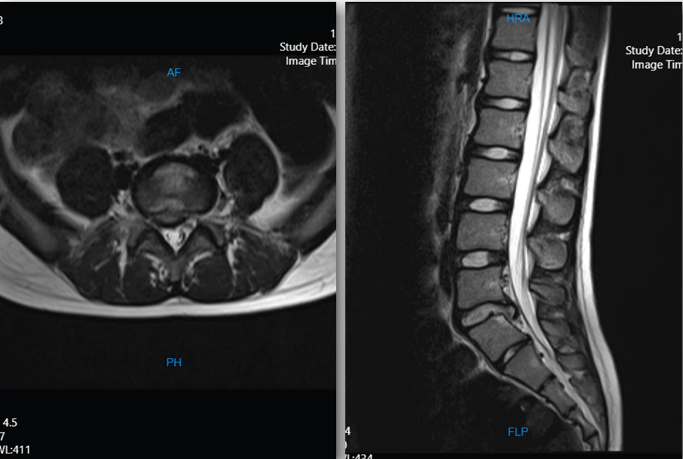
Figure 1: Preoperative MRI of the lumbosacral spine showing right paracentral disc herniation at L5/S1 with foraminal stenosis and caudal fragment migration.
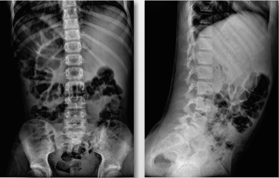
Figure 2: Preoperative AP and lateral plain radiographs of lumbosacral spine showing mild retrolisthesis of L5/S1 without structural anomaly or fracture.
Surgical Technique and Treatment
Conservative management program with analgesics (NSAIDs), relative rest and modification of physical activity was started in the patient but did not achieve satisfactory pain relief. The decision to proceed with surgical intervention was made in light of failure of nonsurgical therapy for greater than four weeks and progressive neurologic deficits.
The patient was taken to the prone position on a Jackson spinal table with appropriate pediatric padding and placed under general endotracheal anesthesia. Routine surgical preparation and draping were performed. A midline incision was made over the L5/S1 level under fluoroscopy. The length of the incision was about 2 cm. A right sided laminotomy and foraminotomy at L5/S1 was performed using microsurgical technique. The traversing S1 nerve root was identified and preserved carefully. A sequestered disc fragment of approximately 1.0 × 1.5 cm was removed from the epidural space and sent for histopathological examination. Haemostasis was carefully performed and the wound closed anatomically in layers. The patient had an uneventful postoperative recovery and was shifted to the ward with complete resolution of the preoperative radicular pain. A postoperative Lumbosacral Orthosis (LSO brace) was prescribed to minimize the risk of spinal instability during recovery. The patient was discharged in a good general condition. 2.4 Pathological Findings Histologic examination of the specimen removed demonstrated fibrocartilaginous disc tissue with mild chronic inflammatory infiltrate that was consistent with herniated nucleus pulposus material. No neoplastic, infectious or granulomatous pathology was present. 2.5 Outcome The patient had complete resolution of right lower extremity radicular pain and paraesthesia at discharge and at outpatient follow-up. Motor strength in right ankle dorsiflexion and plantarflexion was 5/5. The Achilles reflex was bilaterally normal. No postoperative complications were observed, such as wound infection, CSF leak, or neurologic deterioration.
Discourse
Lumbar disc herniation in children is a rare but clinically significant condition. The overall incidence of LDH in children less than 18 years of age is 0.4–15.4% of all surgical cases. However, cases occurring before the age of 12 years are very rare and have different pathophysiological considerations [2,3]. Our patient is a 10-year-old girl, one of the youngest reported cases of surgically managed LDH in the literature, hence the importance of early recognition and adequate management. LDH in children is due to different causes than in adults. In children, trauma and sport injuries are the most common precipitating factors, accounting for approximately 20-30% of cases [6,7,12]. The patient’s history of pain onset after repetitive loading during volleyball activity is consistent with this literature. Structural abnormalities found radiographically in 20-70% of pediatric LDH patients include spondylolisthesis, transitional segmental anomalies, spina bifida occulta, and scoliosis [6,7,13]. In our case, we found mild retrolisthesis at L5/S1 which could have predisposed the disc to herniation under repetitive mechanical stress. Although no formal genetic testing was completed, emerging evidence has linked polymorphisms of genes involved in collagen synthesis (COL9A2, COL11A1) to early onset disc degeneration in pediatric patients [8]. The clinical picture of LDH in children may differ greatly from that in adults. Children often present with lumbar rigidity, scoliotic posture, antalgic gait and hamstring tightness rather than classical radiculopathy [4,5,14]. Bladder and bowel dysfunction is uncommon in children [6]. We found a positive SLR, which has been reported to be positive in up to 90% of pediatric LDH cases, similar to the adult population, motor deficits, and decreased reflexes, all giving a clear neurologic signature that would suggest a surgically important herniation [6,15]. Pediatric LDH distribution is similar to that seen in adults, with L5/S1 being the most common level involved, followed by L4/L5 [6,16,17]. Most series show a slight preponderance of males, although some series of younger pediatric cohorts show a female predominance, consistent with our case [6,16,18]. Clinicians treating this population should be concerned about high-impact sports such as volleyball and gymnastics that seem to put female adolescents at risk for disc herniation.
MRI of the spine is the gold standard for diagnosis of pediatric LDH, providing better visualization of disc morphology, neural element compression, and associated signal changes without radiation exposure [9,19]. MRI in this case showed not only the main L5/S1 herniation, but also the caudally migrated sequestered fragment behind S1. This scenario has implications for surgical planning, as it may require an extended foraminotomy or epidural exploration. Radiographs provide complementary information regarding segmental alignment and structural abnormalities but are not sensitive for soft tissue pathology [9]. Management of pediatric LDH is still a subject of controversy. Treatment is conservative as the first line of treatment, including analgesics, activity restriction for at least four to six weeks and physiotherapy [10,20]. Absolute bed rest is not generally recommended; activity modification is preferred, with strict rest reserved for one to two days in severe cases [20]. The epidural steroid injections and the trigger-point injections are not recommended routinely in the pediatric population due to the lack of supportive evidence and concerns regarding the growth effects [20].
The indications for surgery are: (1) failure of conservative treatment after an adequate trial, (2) significant or progressive neurologic deficits, such as bladder or bowel dysfunction, or (3) disabling pain despite adequate nonsurgical treatment [21,22]. The failure of conservative treatment and progressive motor deficits in our patient were a clear indication for surgery.
The surgical technique is unilateral laminotomy with foraminotomy and microsurgical fragment extraction, the most frequently used technique for pediatric and adult LDH surgery and supported by current evidence of minimal tissue disruption [10,22,23]. Minimally invasive techniques have been reported in some pediatric series with comparable outcomes, less blood loss and shorter hospital stay, including tubular retractor-assisted microdiscectomy and percutaneous endoscopic approaches [24,25]. The choice of technique should be customized depending on the anatomy of herniation, expertise of the surgeon and resources.
Surgical discectomy in children has been associated with favorable short-term results with around 90% of patients experiencing significant pain reduction postoperatively [10,12-14,16]. Two- to three-year follow-up data indicate low rates of symptom recurrence [12,14-16]. Consistent with these good results, our patient had total resolution of radiculopathy and normal neurological exam on discharge.
But the long-term results are less certain. Reoperation rates have been reported from 6% to 24% in various series, and most long-term studies document revision surgery rates of around 20% over a 10-year period [13,14,19]. These results emphasize the importance of long-term structured follow-up and, if possible, the use of minimally invasive techniques to preserve posterior elements of the spine and reduce postoperative instability [22,24].
A curious finding in the pediatric age group is the possibility of spontaneous resolution of symptoms not related to reabsorption of the disc after conservative treatment. Several reports describe asymptomatic patients with persistent radiological herniation on long-term follow-up MRI implying adaptive pain desensitization and dynamic neurological compensation mechanisms distinct from adult pathophysiology [20]. This underscores the reason for a full initial trial of nonsurgical management prior to surgical intervention in neurologically intact patients.
The incidence of spinal disorders in the pediatric population is increasing, partly due to sedentary lifestyles, decreased physical activity, and increased screen time, and begs for targeted preventive strategies. Periodic axial core strengthening exercises and age specific physical activity, especially in children participating in high demand sports, are likely to reduce the burden of sports-related disc injuries in this susceptible population.
Summary
Lumbar disc herniation is uncommon in children, but should be considered in any child with mechanical low back pain and radicular features, especially in the context of trauma related to sports. MRI is the gold standard imaging modality and is crucial in assessing herniation morphology and planning surgical approach. Conservative treatment remains the mainstay in initial management. However, in the presence of neurological deficits or failure of conservative treatment, surgical discectomy provides excellent short-term results. Considering the recurrence and reoperation rates reported in the literature, which are not insignificant, long-term follow-up is mandatory. This case contributes to the limited evidence base on disc herniation in the pre-adolescent age group and highlights the importance of high clinical suspicion, early MRI evaluation, and individualized management.
Declarations Ethical Approval and Consent
Written informed consent was obtained from the patient’s legal guardian for the publication of this case report and any accompanying images. Patient anonymity has been maintained in the manuscript. The ethics committee of Kashan University of Medical Sciences approved this study (IR.TUMS.VCR.REC.1397.070).
Competing Interests: The authors declare no competing interests.
Funding
This work did not receive any specific funding from any funding agency in the public, commercial or not-for-profit sectors.
Authors’ Contributions
KM and MSF contributed equally to data collection, literature review, and manuscript preparation (co-first authors). PY was responsible for case management, performed the surgery and critically reviewed the manuscript for intellectual content. All authors read and approved the final manuscript.
Acknowledgements
The authors thank the patient and her family for permission to publish this case.
References
- Haidar R, Ghanem I, Saad S, Uthman I (2010) Lumbar disc herniation in young children. Acta Paediatr 99: 19-23.
- Lavelle WF, Bianco A, Mason R, Betz RR, Albanese SA (2011) Pediatric disk herniation J Am Acad Orthop Surg 19: 649-656.
- Bradford DS, Garcia A (1969) Herniations of the lumbar intervertebral disk in children and adolescents. A review of 30 surgically treated cases. JAMA 210: 2045-2051.
- Parisini P, Di Silvestre M, Greggi T, Miglietta A, Paderni S (2001) Lumbar disc excision in children and adolescents. Back 26: 1997-2000.
- Strömqvist F, Strömqvist B, Jönsson B, Gerdhem P, Karlsson MK (2016) Lumbar disc herniation surgery in children: outcome and gender differences. Eur Spine J 25: 657-663.
- DeOrio JK, Bianco Jr AJ (1982) Lumbar disc excision in children and adolescents. J Bone Joint Surg Am 64: 991-996.
- Borgesen SE, Vang PS (1974) Herniation of the lumbar intervertebral disk in children and adolescents. Acta Orthop Scand 45: 540-549.
- Rajasekaran S, Kanna RM, Bhargavan R, Shetty AP (2014) Modic changes, disc degeneration and disc herniation in young and adolescent lumbar disc disease. J Spinal Disord Tech 27: E259-E269.
- McAvoy M, McCrea HJ, Chavakula V, Choi H, Bi WL, et al. (2019) Long-term outcomes of lumbar microdiscectomy in the pediatric population: a large single-institution case series. J Neurosurg Pediatr 24: 549-557.
- Ebersold MJ, Quast LM, Bianco AJ (1987) Results of lumbar discectomy in the pediatric patient. J Neurosurg 67: 643-647.
- Garrido E, Humphreys RP, Hendrick EB, Hoffman HJ (1978) Lumbar disc disease in children. Neurosurgery 2: 22-26.
- Grobler LJ, Simmons EH, Barrington TW (1979) Intervertebral disc herniation in the adolescent. Spine 4: 267-278.
- Gennuso R, Humphreys RP, Hoffman HJ, Hendrick EB, Drake JM (1992) Lumbar intervertebral disc disease in the pediatric population. Pediatr Neurosurg 18: 282-286.
- Durham SR, Sun PP, Sutton LN (2000) Surgically treated lumbar disc disease in the pediatric population: an outcome study. J Neurosurg 92: 1-6.
- Shillito Jr J (1996) Pediatric lumbar disc surgery: 20 patients under 15 years of age. Surg Neurol 46: 14-18.
- Kurihara A, Kataoka O (1980) Lumbar disc herniation in children and adolescents. A review of 70 operated cases and their minimum 5-year follow-up studies. Spine (Phila Pa 1976) 5 :443-451.
- Ferrante L, Mastronardi L, Lunardi P, Puzzilli F, Fortuna A (1992) Lumbar disc herniation in teenagers. Eur Spine J 1: 25-28.
- Epstein JA, Lavine LS (1964) Herniated Lumbar Intervertebral Discs in Teen-Age Children. J Neurosurg 21: 1070-1075.
- Key JA (1950) Intervertebral-disc lesions in children and adolescents. J Bone Joint Surg Am 32A: 97-102.
- Weber H (1983) Lumbar disc herniation. A controlled, prospective study with ten years of observation. Spine (Phila Pa 1976) 8: 131-140.
- Mayer HM, Mellerowicz H, Dihlmann SW (1996) Endoscopic discectomy in pediatric and juvenile lumbar disc herniations. J Pediatr Orthop 5: 39-43.
- Payne 3rd WK, Ogilvie JW (1996) Back pain in children and adolescents. Clin Pediatr (Phila) 43: 899-917.
- Sassmannshausen G, Smith BG (2002) Back pain in the young athlete. Clin Sports Med 21: 121-132.
- Zheng C, Wu F, Cai L (2016) Transforaminal percutaneous endoscopic discectomy in the treatment of far-lateral lumbar disc herniations in children. Int Orthop 40: 1099-1102.
- Webb JH, Svien HJ, Kennedy RL (1954) Lumbar intervertebral disk protrusions in children. JAMA 154: 1153-1154.